The man who mapped the human brain: The revolutionary path of a passionate neurologist
🚀 Key Takeaways
- Core concept : Penfield created functional maps of the cortex by stimulating awake patients during epilepsy surgery.
- Practical tip : When learning complex topics, sketch and label regions by function (a simple map helps long-term memory).
- Did you know : The Montreal Neurological Institute, founded in 1934, remains a global center for brain research.
He drew the brain like a cartographer draws a coastline.
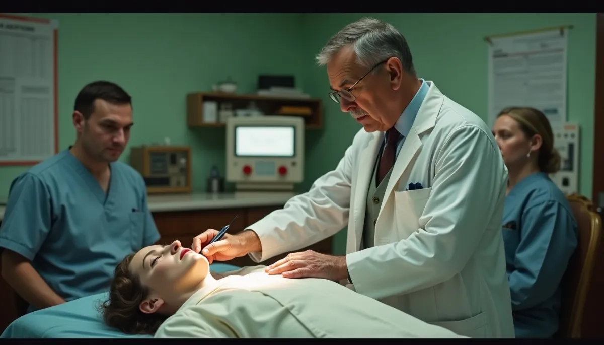
Imagine a dimly lit operating theatre at the Montreal Neurological Institute in the late 1930s. A patient, kept conscious, lies prepared; electrodes hum, a scalp incision opens a landscape of gray and pink. Surgeons and an electrophysiologist watch as tiny currents touch the exposed cortex. The patient reports a twitch, a flash of memory, a sensation in a finger that is not being touched. Each report is a pin on a map, and the map grows, slowly, meticulously.
A living map
Wilder Graves Penfield (1891-1976) was an American-born neurosurgeon who became a towering figure in 20th century neuroscience. In 1934 he founded the Montreal Neurological Institute at McGill University, a center that blended clinical care, research, and teaching.
Between the 1930s and the 1950s, Penfield developed what became known as the Montreal Procedure, an approach to treat drug-resistant epilepsy by removing the epileptic focus while patients were awake. This allowed direct electrical stimulation of the cortex and real-time reports from patients.
From those stimulations emerged the famous cortical homunculus, a distorted human figure representing the amount of cortex dedicated to each body part. That image, first formalized in papers from the late 1930s, remains a visual shorthand in neurology classrooms worldwide.
The surgeon's workshop
Penfield did not work alone. He collaborated closely with colleagues such as Herbert Jasper, an expert in electroencephalography (EEG). Jasper's recordings and Penfield's clinical observations were complementary; together they connected electrical patterns to behavior and sensation.
Concrete milestones anchor this story. The founding of the Montreal Neurological Institute in 1934 created a multidisciplinary hub. In 1937 Penfield published influential mappings of motor and sensory cortex, and across the following decades his team cataloged experiential responses when temporal lobes were stimulated, revealing how stimulation could evoke vivid memories or complex perceptions.
These discoveries had immediate clinical impact. Surgeons gained safer ways to resect epileptic tissue while preserving critical functions. Patients were spared paralysis, language loss, or severe sensory deficits that had been common after earlier operations.
Doubts and horizons
Penfield's findings also provoked deep questions. When stimulation of certain areas produced rich recollections, Penfield speculated on the neural basis of memory and even on mind and soul. His later writings, including popular books, mixed empirical observation and philosophical reflection, which some scientists welcomed and others criticized as overreach.
Decades later, new technologies have both confirmed and complicated Penfield's maps. Functional MRI, diffusion tractography, and the Human Connectome Project (launched in 2009) extend his local, stimulation-based maps into large-scale network maps of connectivity. We now see the brain as both localized and distributed: areas matter, but so do their connections.
Practically, Penfield's approach teaches a persistent lesson: combine careful observation with cross-disciplinary tools. For learners, that means pairing hands-on experience (clinical rounds, lab work) with modern imaging and computational models. For readers curious about brain health, simple actionable advice follows: keep active, learn new skills, and sleep well; these behaviors nurture the networks Penfield's maps point to.
Wilder Penfield's life was not only a sequence of papers and procedures. It was a model of curiosity, precision, and the courage to ask patients to speak while surgeons worked. His maps were born of trust and careful listening, and they continue to guide both operating rooms and research labs.
Thanks for reading, and don't forget, Enjoy Life Moments!